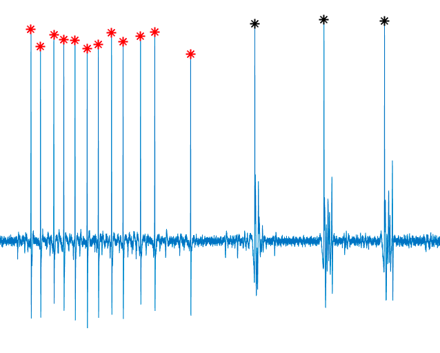
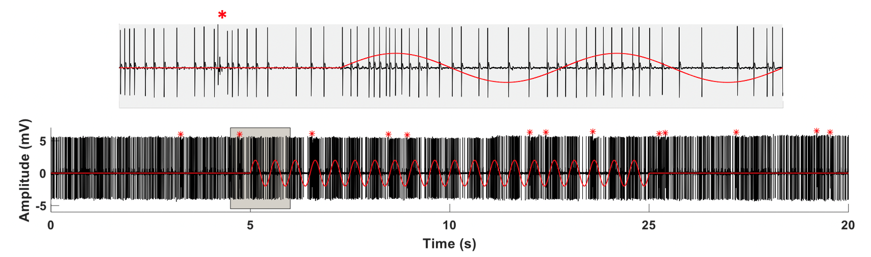
Cerebellar Purkinje cell (PC) simple spikes were entrained by the applied AC field.The effect of applied E-fields was direction and intensity dependent, with rostrocaudally directed fields causing stronger modulations than dorsoventral fields and mediolaterally directed ones causing little to no effect, on average.AC stimulation entrained activity in a frequency dependent manner, with stronger phase-locking to the stimulus cycle at higher frequencies.
Keywords: Cerebellar modulation; Spike entrainment; Transcranial electric stimulation (tES)

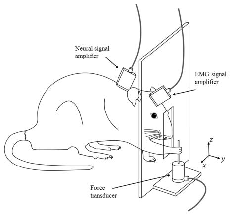

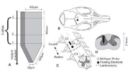
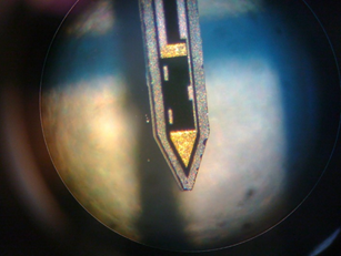
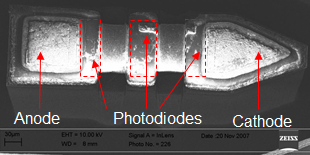
In search of a solution to the long standing problems encountered in traditional brain computer interfaces (BCI), the descending tracts of the spinal cord present an alternative site for taping into the volitional motor signals. Due to the convergence of the cortical outputs into a final common pathway in the descending tracts of the spinal cord, neural interfaces with the spinal cord can potentially acquire signals richer with volitional information in a smaller anatomical region. The main objective of this study was to evaluate the feasibility of extracting motor control signals from the corticospinal tract (CST) of the rat spinal cord. Polyimide substrate, multi-electrode arrays (MEA) were implanted in the CST of rats trained for a lever pressing and reach-to-pull tasks. This novel use of flexible substrate MEAs allowed recording of CST activity in behaving animals for a couple of months. Linear regression, and convolutional neural network methods were applied to the neural signals to reconstruct the isometric forelimb forces first and then the EMG signals. The results support the feasibility of a spinal cord-computer interface with limited but functional level output signals.
Over years this project has been funded by NIH (RO1), The Whitaker Foundation, and The Christopher Reeve Paralysis Foundation.
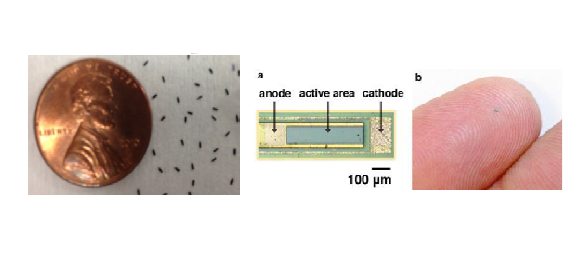
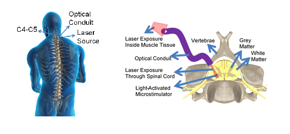
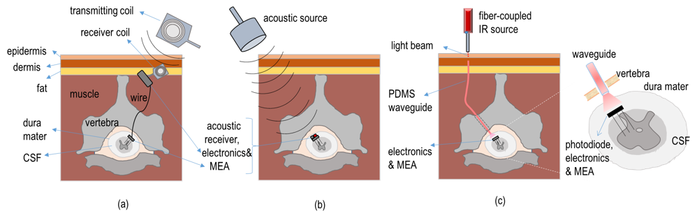
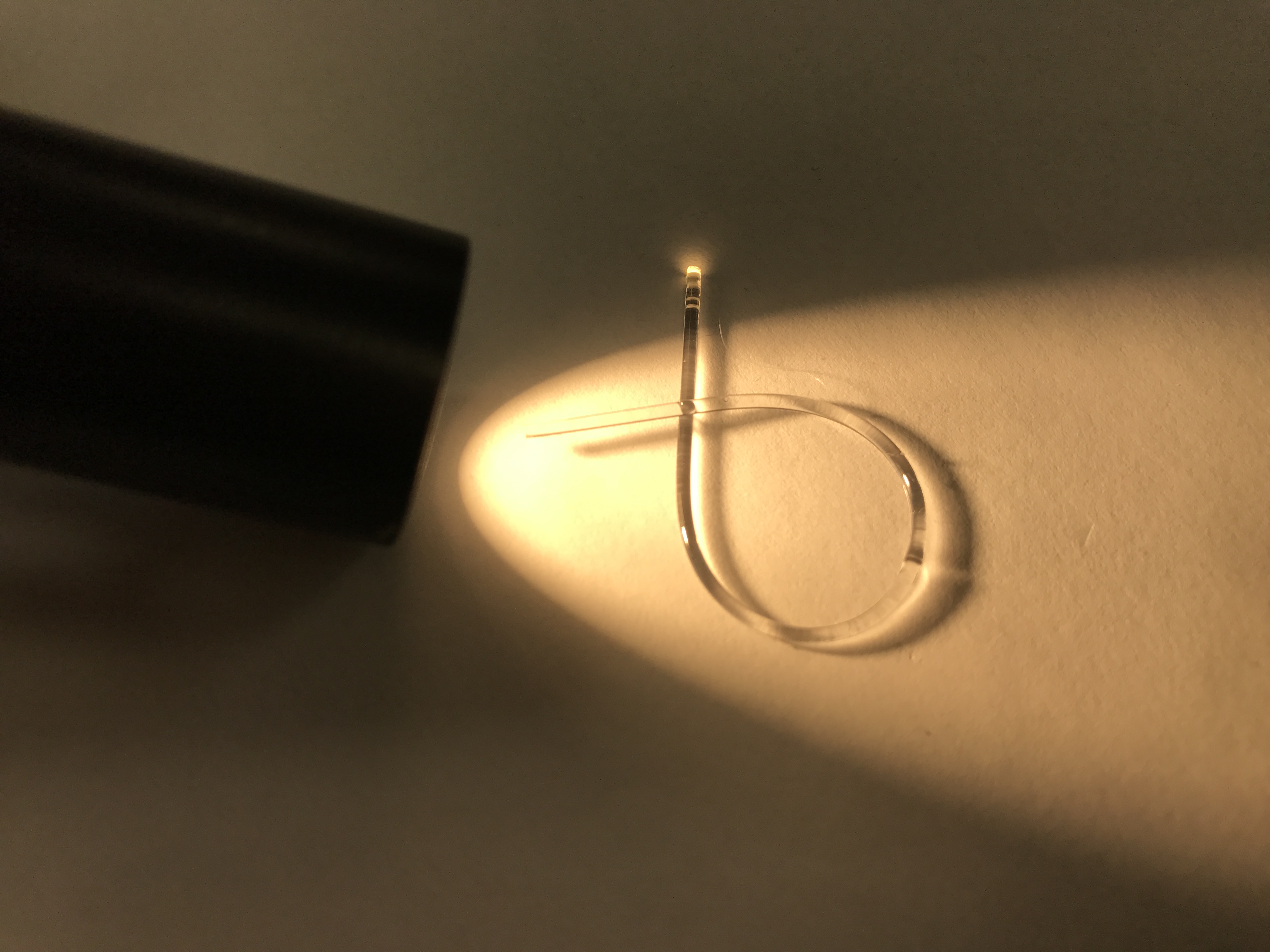
Microelectrode implants often fail either due to the chronic tissue response caused by the tethering forces of the wires or their breakage. Our laboratory is developed a floating light activated micro electric stimulator (FLAMES) that is a wireless implantable device for neural stimulation where near-infrared (NIR) light is used for energy transfer to the microstimulator through neural tissue. The FLAMES devices with sub-millimeter sizes were acutely tested in the rat spinal cord for feasibility of the main concept. Temperature elevation profile was also measured experimentally using a micro termocouple inside the rat brain induced by an NIR laser beam to determine the maximum allowable optical power. Our latest chronic implants have shown minimal tissue response to untethered devices implanted into the brain and the spinal cord in rats. We also developed a PDMS waveguide that can withstand bending and stretching in the body while delivering the optical energy to devices implanted deeper into the tissue such as the spinal cord. These studies have demonstrated the feasibility of microstimulators that can be activated optically by NIR light either by photon penetration through neural tissue (implants near CNS surface) or via optical waveguides (deeper implants) without using any tethering wires. Semiconductor devices were designed and fabricated in collaboration with Dr. Unlu's group at Boston University.
Keywords: optical neural stimulation, near infrared light (NIR), intraspinal micro stimulation
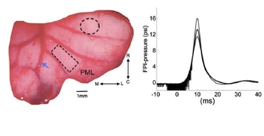
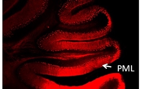
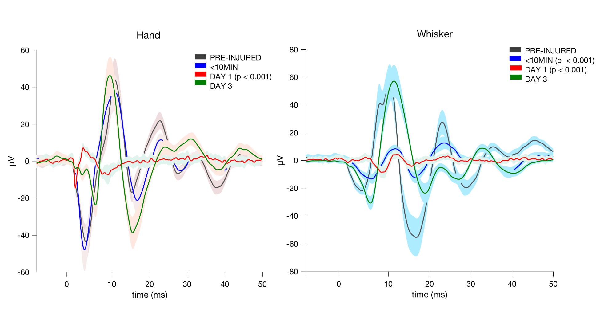
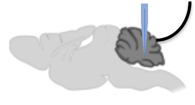
The changes of excitability in affected neural networks can be used as a marker to study the temporal course of traumatic brain injury (TBI). The cerebellum is an ideal platform to study brain injury mechanisms at the network level using the electrophysiological methods. Within its crystalline morphology, the cerebellar cortex contains highly organized topographical subunits that are defined by two main inputs, the climbing (CFs) and mossy fibers (MFs). Here we demonstrate the use of cerebellar evoked potentials (EPs) mediated through these afferent systems for monitoring the injury progression in two different rat models of injury: fluid percussion injury (FPI) and Blast Injury (BI) models. A mechanical tap on the dorsal hand or whiskers were used as a stimulus, and EPs were recorded from the surface of the paramedian lobule (PML) of the posterior cerebellum via flexible multi-electrode arrays (MEAs).
Keywords: traumatic brain injury, evoked potentials, somatotopic organization
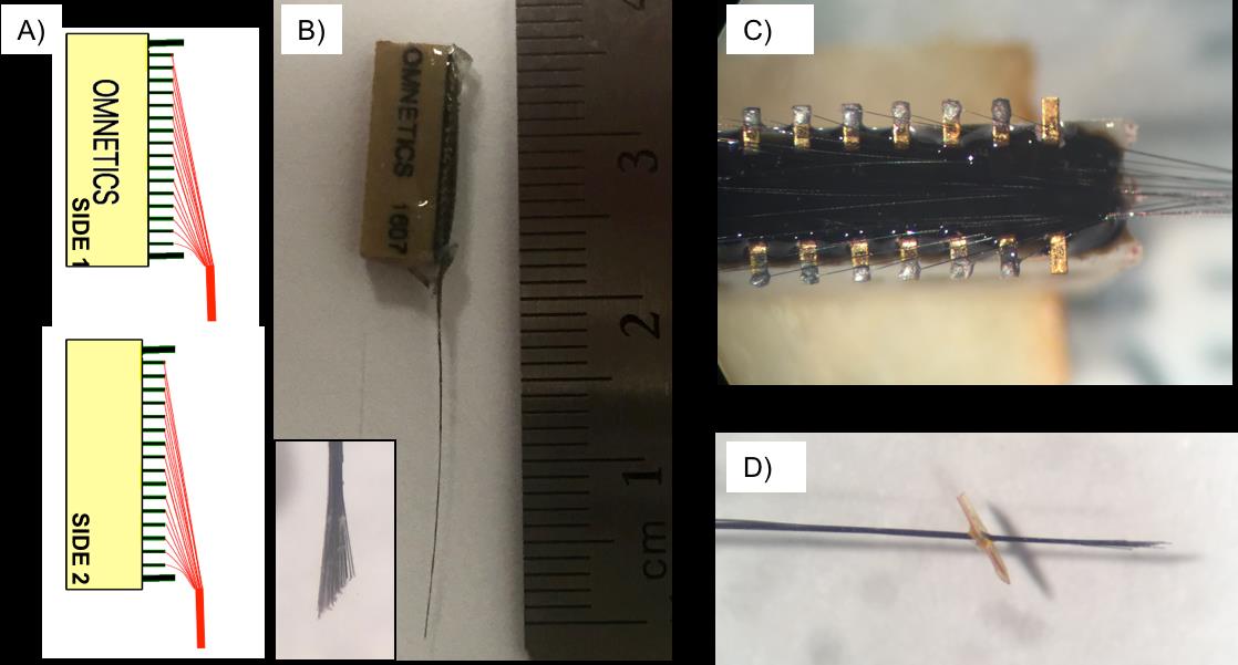
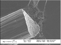
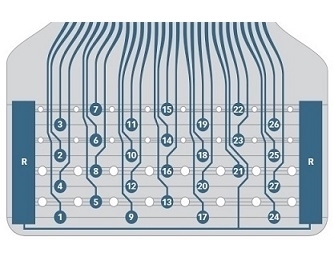
Electrode Fabrication: Parylene-C coated 7 μm carbon fibers were connected to the pins on the Omnetics micro-connector individually using conductive silver epoxy (EpoTek, Massachusetts) and cured in an oven at 150˚C for 10 minutes. The other ends of carbon fibers were individually placed in a 4 by 8 array with 100 μm (vertical) or 150 μm (horizontal) pitch. Electrode impedances were measured in saline at 1 kHz and found to vary from 0.5 to 3 MΩ prior to implantation. We studied sensori-motor content of cerebellar activity using carbon fiber electrode arrays in rats during reach-to-pull tasks. More recently, we used carbon fiber bundle electrodes for recordings and demonstration of neural modulation in the cerebellar nuclei in awake rats during cerebellar transcranial AC stimulation (ctACS).
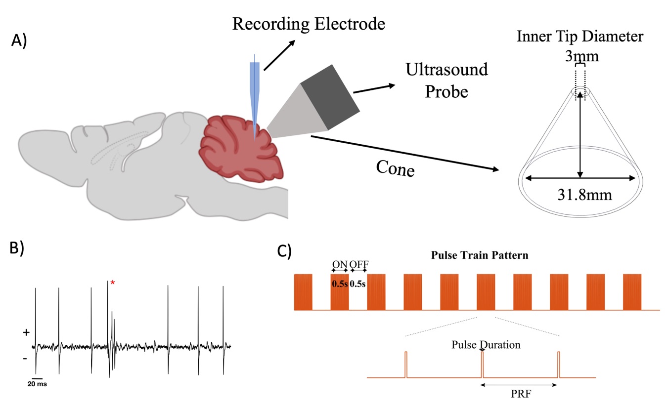
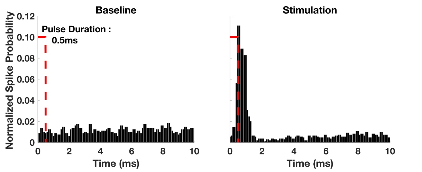
Focused ultrasound (FUS) has excellent characteristics over other non-invasive stimulation methods in terms of spatial resolution and steering capability of the target. FUS has not been tested in the cerebellar cortex and cellular effects of FUS are not fully understood. The objective is to investigate how the activity of cerebellar Purkinje cells (PCs) is modulated by FUS with varying pulse durations and pulse repetition frequencies.
Keywords: Focused ultrasound stimulation; Neuromodulation